Advantages of PandiCath®

Atraumatic

Easy to use and repeat

Independent of anatomic variations

Does not require specialist centers and expertise

Does not cause complications

Prolonged use: from several hours to several days

The only device capable of creating and maintaining isolated area with controlled conditions in GI tract while preserving its connectivity and function


The X-ray image shows radiocontrast fluid injected in the main channel of PandiCath® first accumulating proximally of the upper balloon, with subsequent delivery of the fluid into small intestine, while bypassing the area of duodenum isolated by the balloons.
Placement of PandiCath®
Placement of PandiCath® is a standard endoscopic procedure, which does not require highly specialized skills, equipment or facilities. In fact expanded balloons fix the catheter in place and make PandiCath® installation easier compared to a regular nasojejunal tube, since there is reduced risk of PandiCath® being pulled back when endoscope is removed. The recommended methods of placement of PandiCath® are via a guidewire or with the aid of a guiding thread (see video example of PandiCath® placement below). Correct positioning of the catheter can be easily checked endoscopically, at the end of the placement procedure, and subsequently confirmed radiologically by observing the radiopaque marks located around the balloons or by using radiocontrast solution to inflate the balloons or by injecting radiocontrast solution into the isolated area between the balloons (see X-ray image below) unless counter-indicated.
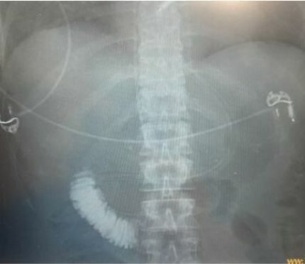
Location of PandiCath® verified by injection of radiocontrast solution into the isolated area between the balloons and X-ray imaging.